
Light Microscopy Tools
Contact
GFZ Websites
Infrastructure belongs to
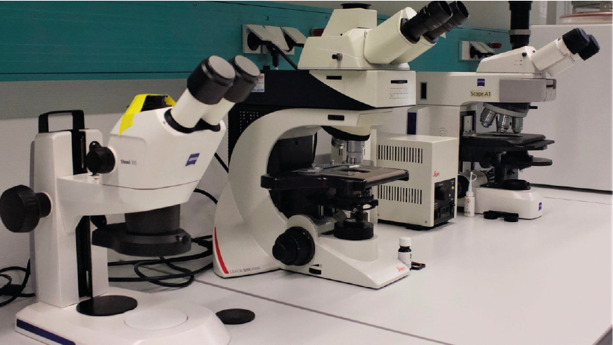
Optical microscopes are available in the Microscopy Lab for different techniques, such as brightfield, polarization contrast, fluorescence. The microscopes can also be equipped with the following cameras: Sony Alpha 6000 digital camera and Kern ODC-86 microscope camera.
The lab is also equipped with a FlowCam 5000 by Yokogawa, which is a particle analyzer capable of taking optical images. It is comparable to a flow cytometer and features a syringe pump and 100 x 700 µm glass capillary, where liquid passes through. During a sample run, images of the flow are taken with a camera, magnified with a 10x objective. Detected particles are compiled as images in a collage, which can then be grouped and classified by the user based on the particle properties (e.g. size description and morphology, among others) provided by the software. Sample volume ranges from 0.25 to 1 mL.
Categories
Disciplinary Keywords
Instrumentation
Laboratory instrumentation
Instruments
-
Fluorescence Microscope
Fluorescence illumination and observation is the most rapidly expanding microscopy technique employed today, both in the medical and biological sciences, a fact which has spurred the development of more sophisticated microscopes and numerous fluorescence accessories. Epi-fluorescence, or incident light fluorescence, has now become the method of choice in many applications and comprises a large part of this tutorial. We have divided the fluorescence section of the primer into several categories to make it easier to organize and download. Please follow the links below to navigate to points of interest. Introductory Concepts - Fluorescence is a member of the ubiquitous luminescence family of processes in which susceptible molecules emit light from electronically excited states created by either a physical (for example, absorption of light), mechanical (friction), or chemical mechanism. Generation of luminescence through excitation of a molecule by ultraviolet or visible light photons is a phenomenon termed photoluminescence, which is formally divided into two categories, fluorescence and phosphorescence, depending upon the electronic configuration of the excited state and the emission pathway. Fluorescence is the property of some atoms and molecules to absorb light at a particular wavelength and to subsequently emit light of longer wavelength after a brief interval, termed the fluorescence lifetime. The process of phosphorescence occurs in a manner similar to fluorescence, but with a much longer excited state lifetime. Anatomy of the Fluorescence Microscope - In contrast to other modes of optical microscopy that are based on macroscopic specimen features, such as phase gradients, light absorption, and birefringence, fluorescence microscopy is capable of imaging the distribution of a single molecular species based solely on the properties of fluorescence emission. Thus, using fluorescence microscopy, the precise location of intracellular components labeled with specific fluorophores can be monitored, as well as their associated diffusion coefficients, transport characteristics, and interactions with other biomolecules. In addition, the dramatic response in fluorescence to localized environmental variables enables the investigation of pH, viscosity, refractive index, ionic concentrations, membrane potential, and solvent polarity in living cells and tissues. Additional information available at: "http://micro.magnet.fsu.edu/primer/techniques/fluorescence/ fluorhome.htm (Source: Global Change Master Directory (GCMD). 2023. GCMD Keywords, Version 16.3. Greenbelt, MD: Earth Science Data and Information System, Earth Science Projects pision, Goddard Space Flight Center (GSFC) National Aeronautics and Space Administration (NASA). URL (GCMD Keyword Forum Page): https://forum.earthdata.nasa.gov/app.php/tag/GCMD+Keywords)
-
Optical Microscope
-
Particle Analyzer
-
Polarization Microscope