
RFA Element-Scanning Labor
Kontakt
GFZ Webseiten
Infrastrukturzugehörigkeit
Das Labor für RFA (Röntgenfluoreszenz)-Element-Scanning konzentriert sich auf zerstörungsfreie und kontinuierliche Kernanalysen. Diese Analysen ermöglichen eine detaillierte geochemische Charakterisierung kompletter Sedimentkerne bis hin zu einzelnen Schichten und Körnern. Die Überbrückung dieser Skalen wird erreicht durch eine Kombination von RFA-Kernscans, Mikro-RFA-Scans von epoxyimprägnierten Sedimentblöcken oder Dünnschliffen, und Rasterelektronenmikroskop-Analysen (REM).
Die Entwicklungen und Forschungen konzentrieren sich auf die Verbesserung von Umwelt- und Klimainterpretationen durch:
-
Integration von RFA-Analysen mit Kern- und Dünnschliffbeobachtungen
-
Geochemische Proxies und Charakterisierung von Sedimenten durch multivariate statistische Analysen
Ausgewählte Infrastrukturen
Fachspezifische Schlagworte
- X-Ray Fluorescence Spectroscopy
- Scanning Electron Microscopy
- X-Ray Fluorescence Core Scanning
- Micro-X-Ray Fluorescence Spectroscopy
- Energy Dispersive Analysis of X-Ray
- Geologie
- Paläoklima
- Sedimentologie
Kategorien
Instrumentierung
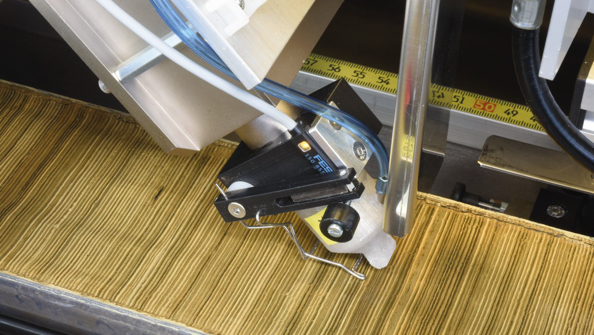
Im Einzelnen ist das Labor mit einem Röntgenfluoreszenz-Kernscanner ausgestattet, um Bohrkern-Halbschalen mit einer Länge von bis zu 180 cm und einem Durchmesser zwischen 6 und 12 cm zu analysieren. Eine CCD-Line-scan-Kamera wird für die optische Linien-Scan-Aufnahme der Bohrkern-Halbschalen mit separaten RGB-Sensoren und für die UV-Lumineszenz-Scans von Karbonatproben (z. B. Speleotheme, Korallen) verwendet. Darüber hinaus wird ein Mikro-Röntgenfluoreszenzscanner zur Messung von Polierte und mit Epoxidharz imprägnierte Sedimentproben, Gesteinsdickschliffe und Speläothemproben (Probenstufe 20 x 17 cm) eingesetzt. Zusätzlich kann ein Desktop-SEM mit EDS für unbeschichtete Proben bis zu einer Länge von 100 mm verwendet werden.
Instrumente
-
X-Ray Fluorescence Core Scanner
-
Micro-X-Ray Fluorescence Spectrometer
Measurement method of X-Ray fluorescence used to measure amounts of elements in a material. Micro-XRF (μ-XRF) analysis uses highly brilliant X-Ray sources (synchrotron source and spot size 100 nm to 2 μm) and microfocussing X-Ray optics to give fg to ag detection limits. (Source: IUPAC; https://doi.org/10.1515/pac-2019-0302)
-
Scanning Electron Microscope
The Scanning Electron Microscope (SEM) is one of the most versatile and widely used tools of modern science as it allows the study of both morphology and composition of biological and physical materials. By scanning an electron probe across a specimen, high resolution images of the morphology or topography of a specimen, with great depth of field, at very low or very high magnifications can be obtained. Compositional analysis of a material may also be obtained by monitoring secondary X-rays produced by the electron-specimen interaction. Thus detailed maps of elemental distribution can be produced from multi-phase materials or complex, bio-active materials. Characterization of fine particulate matter in terms of size, shape, and distribution as well as statistical analyses of these parameters, may be performed. There are many different types of SEM designed for specific purposes ranging from routine morphological studies, to high-speed compositional analyses or to the study of environment-sensitive materials. The Centre for Microscopy & Microanalysis presents three particular types of SEM that, in combination, provide a powerful analytical approach for many research or quality-control applications. Additional information available at "http://www.uq.edu.au/nanoworld/sem_gen.html" [Summary provided by The University of Queensland] (Source: Global Change Master Directory (GCMD). 2023. GCMD Keywords, Version 16.3. Greenbelt, MD: Earth Science Data and Information System, Earth Science Projects pision, Goddard Space Flight Center (GSFC) National Aeronautics and Space Administration (NASA). URL (GCMD Keyword Forum Page): https://forum.earthdata.nasa.gov/app.php/tag/GCMD+Keywords)